Medicine has seen tremendous innovations, yet ultrasound remains a mainstay for its safety and versatility. The Endocavitary Probe elevates this technology by positioning the ultrasound source inside the body, enabling sharper imaging and real-time procedural assistance that redefine clinical diagnostics and interventions.
Engineering Insights into the Endocavitary Probe
Acoustic Design Features
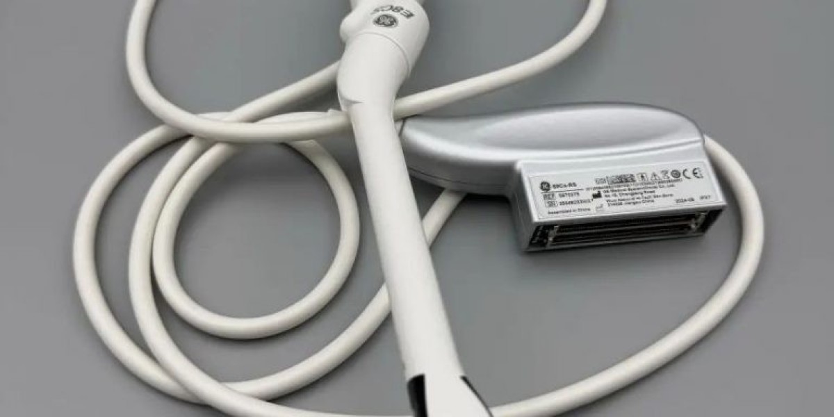
At the heart of the Endocavitary Probe lies a high-density piezo crystal array. Its small footprint and variable curvature facilitate consistent contact with irregular tissue surfaces, improving acoustic coupling and image fidelity.
Integrated Ergonomics
Cable strain relief, intuitive controls, and angle-adjustable necks make these probes more user-friendly. Sonographers and physicians can maneuver them easily, even in tight anatomical spaces.
Full-Spectrum Clinical Deployment
Women’s Health Applications
For gynecologists, transvaginal Endocavitary Probes are essential tools. They reveal corpus luteum, submucosal fibroids, and early embryo developments—guiding fertility treatments and early pregnancy monitoring.
Prostate and Rectal Pathology
Transrectal probes assist urologists in visualizing prostate size, detecting nodules, and performing ultrasound-guided biopsies. They also aid colorectal specialists in evaluating rectal wall lesions with high-resolution detail.
Cardiac & Thoracic Adjunct
Transesophageal echo (TEE) probes are a subtype of Endocavitary Probe, used extensively in cardiac surgery to assess valves and intracardiac structures in real time—especially during procedures involving anesthesia or open-heart interventions.
Clinical Outcomes Enhancement
Accelerated Diagnosis
Immediate visualization allows rapid triage. Small cysts, polyps, or suspect tissue can be observed and acted upon in the same session—eliminating delays and follow-up visits.
Less Invasive yet Precise
Minimizing the need for incisions, the Endocavitary Probe supports minimally invasive processes with maximal diagnostic precision, resulting in fewer complications and accelerated recovery.
Monitoring Treatment Progress
In fertility medicine or cancer therapy, these probes monitor treatment efficacy, measuring structural changes and blood flow alterations—providing crucial data for clinical decisions.
Navigating Challenges
Overcoming Discomfort
Some patients may experience discomfort or anxiety. Using topical anesthetics, offering sedatives in certain cases, and allowing for shorter scan durations can make the process more tolerable.
Technical Limitations
High-frequency probes have limited penetration depth. This means they’re ideal for superficial structures within cavities but less so for deeper masses—another reason for complementary imaging in select cases.
Regulatory and Safety Standards
Medical boards require protocols for periodic sterilization checks, probe integrity tests, and documentation. Compliance ensures patient protection and legal adherence.
Conclusion
By embracing inside-out imaging, the Endocavitary Probe has reshaped clinical workflows. It delivers immediate, high-resolution visuals where they matter most—inside cavities—boosting diagnostic confidence and care efficiency. With robust sterilization protocols and awareness of limitations, it stands as a leading tool in modern medicine’s toolkit.